What is Arachnoid Cysts?
Arachnoid cysts are cerebrospinal fluid covered by arachnoidal cells and collagen that may develop between the surface of the brain and the cranial base or on the arachnoid membrane, one of the three meningeal layers that cover the brain and the spinal cord. Arachnoid cysts are a congenital disorder, and most cases begin during infancy; however, onset may be delayed until adolescence.
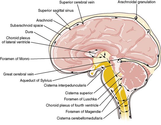
Arachnoid cysts are relatively common benign and asymptomatic lesions occurring in association with the central nervous system, both within the intracranial compartment (most common) as well as within the spinal canal. They are usually located within the subarachnoid space and contain CSF.
On imaging, they are characterised as well circumscribed cysts, with an imperceptible wall, displacing adjacent structures, and following the CSF pattern (hypodense on CT and hyperintense on T2 with FLAIR suppression on MRI). They can also have a remodelling effect on the adjacent bone.
Arachnoid cysts are cerebrospinal fluid-filled sacs that are located between the brain or spinal cord and the arachnoid membrane, one of the three membranes that cover the brain and spinal cord. Primary arachnoid cysts are present at birth and are the result of developmental abnormalities in the brain and spinal cord that arise during the early weeks of gestation.
Secondary arachnoid cysts are not as common as primary cysts and develop as a result of head injury, meningitis, or tumors, or as a complication of brain surgery. The majority of arachnoid cysts form outside the temporal lobe of the brain in an area of the skull known as the middle cranial fossa. Arachnoid cysts involving the spinal cord are rarer. The location and size of the cyst determine the symptoms and when those symptoms begin. Most individuals with arachnoid cysts develop symptoms before the age of 20, and especially during the first year of life, but some people with arachnoid cysts never have symptoms. Males are four times more likely to have arachnoid cysts than females.
Typical symptoms of an arachnoid cyst around the brain include headache, nausea and vomiting, seizures, hearing and visual disturbances, vertigo, and difficulties with balance and walking. Arachnoid cysts around the spinal cord compress the spinal cord or nerve roots and cause symptoms such as progressive back and leg pain and tingling or numbness in the legs or arms. Diagnosis usually involves a brain scan or spine scan using diffusion-weighted MRI (magnetic resonance imaging) which helps distinguish fluid-filled arachnoid cysts from other types of cysts.
Arachnoid cysts are benign cysts that occur in the cerebrospinal axis in relation to the arachnoid membrane and that do not communicate with the ventricular system. They usually contain clear, colorless fluid that is most likely normal cerebrospinal fluid; rarely, they contain xanthochromic fluid.
Most are developmental anomalies. A small number of arachnoid cysts are acquired, such as those occurring in association with neoplasms or those resulting from adhesions occurring in association with leptomeningitis, hemorrhage, or surgery
Arachnoid cysts are cerebrospinal fluid-filled sacs that are located between the brain or spinal cord and the arachnoid membrane, one of the three membranes that cover the brain and spinal cord. Primary arachnoid cysts are present at birth and are the result of developmental abnormalities in the brain and spinal cord that arise during the early weeks of gestation. Secondary arachnoid cysts are not as common as primary cysts and develop as a result of head injury, meningitis, or tumors, or as a complication of brain surgery.
The majority of arachnoid cysts form outside the temporal lobe of the brain in an area of the skull known as the middle cranial fossa. Arachnoid cysts involving the spinal cord are rarer. The location and size of the cyst determine the symptoms and when those symptoms begin. Most individuals with arachnoid cysts develop symptoms before the age of 20, and especially during the first year of life, but some people with arachnoid cysts never have symptoms. Males are four times more likely to have arachnoid cysts than females.
Typical symptoms of an arachnoid cyst around the brain include headache, nausea and vomiting, seizures, hearing and visual disturbances, vertigo, and difficulties with balance and walking. Arachnoid cysts around the spinal cord compress the spinal cord or nerve roots and cause symptoms such as progressive back and leg pain and tingling or numbness in the legs or arms. Diagnosis usually involves a brain scan or spine scan using diffusion-weighted MRI (magnetic resonance imaging) which helps distinguish fluid-filled arachnoid cysts from other types of cysts.
An arachnoid cyst is most likely to develop in your head, but it can also develop around your spinal cord. It’s called an arachnoid cyst because it occurs in the space between your brain, or spinal column, and your arachnoid membrane. This is one of three membrane layers that surround your brain and spine. If an arachnoid cyst develops in your head, it will grow between your brain and skull or in pockets around your brain called ventricles.
Arachnoid cysts are usually filled with cerebrospinal fluid (CSF). CSF is a naturally occurring protective fluid that surrounds your brain and spinal column. The walls of an arachnoid cyst don’t allow this fluid to drain into your CSF system, causing it to accumulate inside.
In children, arachnoid cysts are usually congenital, or present at birth. These cysts are called primary arachnoid cysts. Arachnoid cysts that develop later in life are called secondary arachnoid cysts. Primary arachnoid cysts are more common than secondary arachnoid cysts.
Signs and symptoms
Patients with arachnoid cysts may never show symptoms, even in some cases where the cyst is large. Therefore, while the presence of symptoms may provoke further clinical investigation, symptoms independent of further data cannot—and should not—be interpreted as evidence of a cyst’s existence, size, location, or potential functional impact on the patient.
Symptoms vary by the size and location of the cyst(s), though small cysts usually have no symptoms and are discovered only incidentally. On the other hand, a number of symptoms may result from large cysts:
- Cranial deformation or macrocephaly (enlargement of the head), particularly in children
- Cysts in the suprasellar region in children have presented as bobbing and nodding of the head called bobble-head doll syndrome.
- Cysts in the left middle cranial fossa have been associated with ADHD in a study on affected children.
- [2] A patient experiencing a headache does not necessarily have an arachnoid cyst.
- In a 2002 study involving 78 patients with a migraine or tension-type headache, CT scans showed abnormalities in over a third of the patients, though arachnoid cysts only accounted for 2.6% of patients in this study.
- A study found 18% of patients with intracranial arachnoid cysts had non-specific headaches. The cyst was in the temporal location in 75% of these cases.
- Seizures
- Hydrocephalus (excessive accumulation of cerebrospinal fluid)
- Increased intracranial pressure
- Developmental delay
- Behavioral changes
- Nausea
- Hemiparesis (weakness or paralysis on one side of the body)
- Ataxia (lack of muscle control)
- Musical hallucination
- Pre-senile dementia, a condition often associated with Alzheimer’s disease
- In elderly patients (>80 years old) symptoms were similar to chronic subdural hematoma or normal pressure hydrocephalus
- Dementia
- Urinary incontinence
- Hemiparesis
- Headache
- Seizures
Arachnoid cysts are usually asymptomatic. That means they don’t tend to produce symptoms. As a result, most people who have an arachnoid cyst don’t realize it until they’re examined for other issues, such as head injuries.
In some cases, arachnoid cysts do cause symptoms. Symptoms depend on the location and size of the cyst. For example, symptoms may develop if you have a cyst that presses on nerves or sensitive areas of your brain or spinal cord. If it’s located in your brain, the cyst may produce one or more of the following symptoms:
- headache
- dizziness
- nausea
- vomiting
- lethargy
- seizures
- problems hearing, seeing, or walking
- balance issues
- developmental delay
- dementia
If it’s located in your spinal column, it may cause symptoms such as:
- back pain
- scoliosis
- muscle weakness or spasms
- lack of feeling or tingling in your arms or legs
- problems controlling your bladder or bowels
- If you develop these symptoms, contact your doctor.
Causes
The exact cause of arachnoid cysts is not known. Researchers believe that most cases of arachnoid cysts are developmental malformations that arise from the unexplained splitting or tearing of the arachnoid membrane.
In some cases, arachnoid cysts occurring in the middle fossa are accompanied by underdevelopment (hypoplasia) or compression of the temporal lobe. The exact role that temporal lobe abnormalities play in the development of middle fossa arachnoid cysts is unknown.
There are some cases where hereditary disorders have been connected with arachnoid cysts.
Some complications of arachnoid cysts can occur when a cyst is damaged because of minor head trauma. Trauma can cause the fluid within a cyst to leak into other areas (e.g., subarachnoid space). Blood vessels on the surface of a cyst may tear and bleed into the cyst (intracystic hemorrhage), increasing its size. If a blood vessel bleeds on the outside of a cyst, a collection of blood (hematoma) may result. In the cases of intracystic hemorrhage and hematoma, the individual may have symptoms of increased pressure within the cranium and signs of compression of nearby nerve (neural) tissue.
Some scientists debate whether arachnoid cysts are a true congenite condition or if this should be separated from secondary cysts. A recent study shows differences in communication between the arachnoid cyst and the subarachnoid space by CT cisternography . A comparison of arachnoid cyst fluid and CSF in a series of patients show differences in chemical composition.
Arachnoid cysts can also occur secondary to other disorders such as Marfan syndrome, arachnoiditis, or agenesis of the corpus callosum.
Primary, or congenital, arachnoid cysts are usually caused by an abnormal growth of your brain and spinal column while you’re developing in utero. The exact cause of this growth is not known. It might be genetic.
Secondary arachnoid cysts, also known as noncongenital arachnoid cysts, can be caused by several things. These include:
- injury to your head or spinal cord
- complications from brain or spinal surgery
- meningitis
- tumors
Arachnoid cysts are most common in children. They’re more likely to develop in men than women, reports the National Organization for Rare Disorders.
Diagnosis
Diagnosis is principally by MRI. Frequently, arachnoid cysts are incidental findings on MRI scans performed for other clinical reasons. In practice, diagnosis of symptomatic arachnoid cysts requires symptoms to be present, and many with the disorder never develop symptoms.
Additional clinical assessment tools that can be useful in evaluating a patient with arachnoid cysts include the mini-mental state examination (MMSE), a brief questionnaire-based test used to assess cognition.[11]
General imaging differential considerations include:
- enlarged CSF space (e.g. mega cisterna magna)
- epidermoid cyst
- often shows a heterogeneous/dirty signal on FLAIR
- restricted diffusion
- more lobulated
- tend to engulf adjacent arteries and cranial nerves
- subdural hygroma/chronic subdural haemorrhage
- do not typically show CSF signal intensity on MRI
- can have an enhancing membrane
- cystic tumours: often will have a solid/enhancing component and be intra-axial
- pilocytic astrocytoma
- haemangioblastoma
- non-neoplastic cysts
- neurenteric cyst
- neuroglial cyst
- porencephalic cyst
- often follow a history of trauma or stroke
- surrounded by gliotic brain
- neurocysticercosis
- small cyst
- usually multiple when in the subarachnoid space
Untreated, arachnoid cysts may cause permanent severe neurological damage when progressive expansion of the cyst(s) or bleeding into the cyst injures the brain or spinal cord. Symptoms usually resolve or improve with treatment.
If your doctor suspects you have an arachnoid cyst, they will likely order imaging tests. For example, they may use CT and MRI scans to look at your brain or spine.
Treatment
Most arachnoid cysts are asymptomatic and do not require treatment. Treatment may be necessary when symptomatic.[2] A variety of procedures may be used to decompress (remove pressure
- Surgical placement of a cerebral shunt.
- An internal shunt drains into the subdural compartment.
- A cystoperitoneal shunt drains to the peritoneal cavity.
- Fenestration:
- Craniotomy with excision
- Various endoscopic techniques are proving effective,including laser-assisted techniques.
- Drainage by needle aspiration or burr hole.
- Capsular resection
- Pharmacological treatments may address specific symptoms such as seizures or pain.
Arachnoid cysts are benign, and the vast majority remain asymptomatic throughout life. If they are deemed to be causing symptoms, then surgery can be contemplated. This can either take the form of a craniotomy (fenestration or excision) or placement of a cystoperitoneal shunt.
There has been active debate about how to treat arachnoid cysts. The need for treatment depends mostly upon the location and size of the cyst. If the cyst is small, not disturbing surrounding tissue, and not causing symptoms, some doctors will refrain from treatment. In the past, doctors placed shunts in the cyst to drain its fluid. Now with microneurosurgical techniques and endoscopic tools that allow for minimally invasive surgery, more doctors are opting to surgically remove the membranes of the cyst or open the cyst so its fluid can drain into the cerebrospinal fluid and be absorbed.
There has been active debate about how to treat arachnoid cysts. The need for treatment depends mostly upon the location and size of the cyst. If the cyst is small, not disturbing surrounding tissue, and not causing symptoms, some doctors will refrain from treatment. In the past, doctors placed shunts in the cyst to drain its fluid. Now with microneurosurgical techniques and endoscopic tools that allow for minimally invasive surgery, more doctors are opting to surgically remove the membranes of the cyst or open the cyst so its fluid can drain into the cerebrospinal fluid and be absorbed.
If you have an arachnoid cyst that isn’t causing any symptoms or other complications, your doctor may advise you to leave it untreated. They will likely monitor the cyst over time to watch for potential growth or other changes. If it begins to cause problems, they may recommend treatment.
If you have an arachnoid cyst in your brain that’s causing symptoms, your doctor will likely drain it. They may recommend one of two procedures. In the first procedure, they will make a small incision near the cyst and insert an endoscope with a small camera on the end.
They will use this endoscope to gently open the cyst, allowing the fluid inside to drain into your CSF system, where it will be redistributed through your body. This procedure is called fenestration. In the second procedure, your doctor will insert a shunt into the cyst. This will allow the fluid inside to drain to another part of your body, such as your belly.
If you have a symptomatic cyst in your spinal column, your doctor may recommend surgery to remove it completely. If that’s not feasible, they may use fenestration or shunting to drain it.
0 200
No Comments