
Abercrombies syndrome is infiltration of amyloid between cells and fibers of tissues and organs. Amyloid is a waxy protein containing starch and cellulose. This protein is insoluble, which means that it cannot be dissolved or broken down. When amyloid penetrates an organ, it will usually become deposited in the connective tissue cells and the capillary walls.
This condition occurs when amyloid permeates the fibers or cells of a tissue and causes degeneration. Abercrombies syndrome most commonly occurs in the spleen, kidneys and liver, though it can affect any tissue. As the tissue continues to degenerate, it will lose some or all of its normal functioning. Since the symptoms of Abercrombie disease are often vague, this condition might not be diagnosed until a significant amount of degeneration has occurred.
The disease is not an independent one but comes on in certain cachectic states due to chronic tuberculosis, syphilis, diseases of bone involving prolonged suppuration, chronic dysentery, etc.
As more unusual causes of Abercrombies syndrome, may be mentioned leukemia, Hodgkin’s disease (malignant lymphoma), very rarely cancer or sarcoma. It is frequently associated with the kidney with chronic inflammation of that organ, but it is doubtful whether the latter is to be regarded as its cause, for, on the one hand, amyloid disease may lead to nephritis, and, on the other, both conditions may be the result of syphilis.
It is more difficult to determine the nature of the connection between the vice in the blood and the disease in the tissues. By some it is supposed that the, amyloid substance arises in the blood by modification of the albumen and is then infiltrated into the structures.
But this view cannot be accepted, for various reasons. In the first place, the substance is eminently insoluble, and it is difficult to understand how it can be carried by the blood; besides this it does not displace the normal structures simply, but replaces them, these structures being converted into the amyloid substance. It is more consistent to suppose that the tissues are reduced in vitality by the altered condition of the blood, and that the albumen of the blood enters into combination with the protoplasm in such a way as to produce this peculiar substance.
The process may perhaps be compared to the coagulation of the tissues, which, as we have seen, sometimes occurs when they undergo necrosis, the tissues entering apparently into a chemical union with the fibrinogen in the fluid exuded from the blood-vessels, so as to form fibrine or some substance allied to it.
Amyloid matter has frequently been compared to fibrine, and researchers has suggested its affinity with de-alkalized fibrine. The existence of localized amyloid disease is strongly confirmatory of this view. In this condition abnormal structures enter into this peculiar chemical combination with the albumen of the blood, while normal structures do not.
In this connection also, the fact that Abercrombie’s syndrome affects the connective structures of the body is not to be forgotten. It is as if the chemical basis of these structures had a special relation to the amyloid substance. Abercrombie’s syndrome is therefore essentially a degeneration although, in order to the formation of the amyloid substance, it is necessary to have, added to the tissue, material from without, and this adds greatly to the bulk and weight of the structures.
Primary degeneration of the conjunctiva of amyloid nature is a rare but well established condition. Independent studies have been contributed by researchers. A few reports have also appeared from India.
Amyloid degeneration of the conjunctiva has been described in two groups by i) Primary, ii) Secondary. The features of the primary type being the absence of antecedent or co-existant disease, involvement of mesodermal tissues and variability of the staining reaction.
Those of the secondary type are tendency for nodular deposition, consistency in staining reaction of the amyloid material and involvement of the organs like spleen, kidney and liver. In most of the cases reported amyloid degeneration followed some chronic inflammation like trachoma. In all these reported cases bulbar conjunctiva seems to have been unaffected.
The etiology of amyloid degeneration in ocular tissues is unknown. Trachoma is said to be an important cause. Researchers suggested nutritional deficiency as a possible cause. Researchers considered amyloid degeneration as a subgroup of hyaline infiltration.
Symptoms
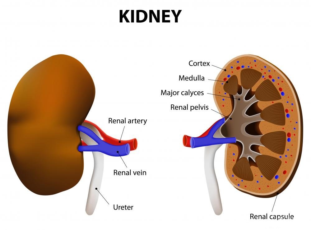
Organs affected by Abercrombies syndrome will typically become enlarged, smooth and hard. The tissue will take on a slightly white or yellow translucent appearance, similar to a bacon rind. Within the tissue, the cortex will also be bloodless. When the blood vessels or muscular overlay of an artery are affected, the tissue will thicken and become transparent. Commonly affected organs include the spleen, kidneys, pancreas and liver. Almost any organ or bodily tissue, however, can become affected by Abercrombie disease.
The symptoms of Abercrombies syndrome vary according to the tissue or organ affected. Since sufferers of this condition are commonly suffering from other wasting diseases, a person’s symptoms might be overlooked. People suffering from Abercrombie syndrome of the kidneys may notice increased urine production, vomiting, diarrhea, bad breath, and edema. Those suffering from degeneration in other organs might notice similar symptoms or even symptoms more specific to the organ’s function.
Diagnosis
The presence of amyloid substance is determined by its physical characters and by certain color tests. The earliest known of this latter is the reaction with iodine. The iodine reaction is useful for roughly testing macroscopically at the time of the post-mortem. For this purpose a watery solution, consisting of iodine 10 grains, iodide of potassium 20 grains, and water 4 ounces, is poured on the surface of the structure to be tested. A mahogany-red color indicates the presence of amyloid matter. The further addition of dilute sulphuric acid sometimes produces a deeper red or a bluish color.
For microscopic purposes watery solutions of methylviolet or gentian violet as introduced by Cornil are most suitable. These dyes produce a rose-pink color with the amyloid substance, while normal tissues are stained blue. In testing by iodine microscopically a solution half the strength of that mentioned above is to be used.
The substance itself has a peculiar bright translucent glancing appearance, and, as the structures in which it occurs are enlarged, they are often remarkably prominent under the microscope. It is a very dense heavy material, and, after death, at least, is somewhat brittle, but the usual absence of hemorrhage in amyloid organs would seem to indicate that it is not so during life.
Blood and urine tests can reveal the presence of amyloid protein, but tissue or bone-marrow biopsy is necessary to positively diagnose amyloidosis. Once the diagnosis has been confirmed, additional laboratory tests and imaging procedures are performed to determine:
which type of amyloid protein is involved
which organ(s) or system(s) have been affected
how far the disease has progressed
Treatment
The treatment must be directed to the improvement of the blood, and to the rapid elimination of all septic material. The anemia which forms a conspicuous part of this disease must be treated with iron and other direct restoratives in order to facilitate the carrying of oxygen into the blood. The use of one or more of our excellent vegetable alternatives will produce favorable results. Echinacea stands at the head and with this there is phytolacca, polymnia, stillingia, baptisia, corydalis, yellow dock, burdock, and dandelion, with the specific liver remedies. These may be persisted in for weeks at a time with only good results. For their general tonic effect the glycerophosphates, hydrastis and strychnin will be selected.
Patients might also be advised to consume a nutritious diet and get regular exercise to inhibit further degeneration.
Prognosis
Most cases of amyloidosis are diagnosed after the disease has reached an advanced stage. The course of each patient’s illness is unique but death, usually a result of heart disease or kidney failure, generally occurs within a few years. Amyloidosis associated by multiple myeloma usually has a poor prognosis. Most patients with both diseases die within one to two years.
Prevention
Genetic couseling may be helpful for patients with hereditary amyloidosis and their families. Use of Cholchicine in patients with familial Mediterranean fever has successfully prevented amyloidosis.
Types of Amyloidosis
The major forms of this disease are primary systemic, secondary, and familial or hereditary amyloidosis. There is also another form of amyloidosis associated with Alzheimer’s disease.
Primary systemic amyloidosis usually develops between the ages of 50 and 60. With about 2,000 new cases diagnosed annually, primary systemic amyloidosis is the most common form of this disease in the United States. Also known as light-chain-related amyloidosis, it may also occur in association with multiple myeloma (bone marrow cancer).
Secondary amyloidosis is a result of chronic infection or inflammatory disease.
It is often associated with:
- familial Mediterranean fever (a bacterial infection characterized by chills, weakness, headache, and recurring fever)
- granulomatous ileitis (inflammation of the small intestine)
- Hodgkin’s disease (cancer of the lymphatic system)
- leprosy
- osteomyelitits (bacterial infection of bone and bone marrow)
- rheumatoid arthritis
Familial or hereditary amyloidosis is the only inherited form of the disease. It occurs in members of most ethnic groups, and each family has a distinctive pattern of symptoms and organ involvement. Hereditary amyloidosis is though to be autosomal dominant, which means that only one copy of the defective gene is necessary to cause the disease. A child of a parent with familial amyloidosis has a 50-50 chance of developing the disease.
Amyloidosis can involve any organ or system in the body. The heart, kidneys, gastrointestinal system, and nervous system are affected most often. Other common sites of amyloid accumulation include the brain, joints, liver, spleen, pancreas, respiratory system, and skin.
For more information visit us our website: https://www.healthinfi.com
0 200
1 Comment