About Arteriovenous Malformation
Introduction
Normally, arteries carry blood containing oxygen from the heart to the brain, and veins carry blood with less oxygen away from the brain and back to the heart. When an arteriovenous malformation (AVM) occurs, a tangle of blood vessels in the brain or on its surface bypasses normal brain tissue and directly diverts blood from the arteries to the veins.
A brain arteriovenous malformation (AVM) is a tangle of abnormal blood vessels connecting arteries and veins in the brain.
The arteries are responsible for taking oxygen-rich blood from the heart to the brain. Veins carry the oxygen-depleted blood back to the lungs and heart. A brain AVM disrupts this vital process. An arteriovenous malformation can develop anywhere in your body but occurs most often in the brain or spine. Even so, brain AVMs are rare and affect less than 1 percent of the population.
The cause of AVMs is not clear. Most people are born with them, but they can occasionally form later in life. They are rarely passed down among families genetically. Some people with brain AVMs experience signs and symptoms, such as headache or seizures. AVMs are commonly found after a brain scan for another health issue or after the blood vessels rupture and cause bleeding in the brain (hemorrhage). Once diagnosed, a brain AVM can often be treated successfully to prevent complications, such as brain damage or stroke.
Find out why Mayo Clinic is the best place for your care.
Arteriovenous malformation (AVM) is an abnormal connection between arteries and veins, bypassing the capillary system. This vascular anomaly is widely known because of its occurrence in the central nervous system (usually cerebral AVM), but can appear in any location. Although many AVMs are asymptomatic, they can cause intense pain or bleeding or lead to other serious medical problems.
AVMs are usually congenital and belong to the RASopathies. The genetic transmission patterns of AVM, if any, are unknown. AVM is not generally thought to be an inherited disorder, unless in the context of a specific hereditary syndrome.
Arteriovenous malformations (AVMs) are congenital lesions composed of a complex tangle of arteries and veins connected by one or more fistulae (see the image below). They most commonly occur in young adults, with morbidity and death occurring in 30–50% and 10–15% of patients, respectively. AVM stands for Arteriovenous Malformation. An AVM is a tangle of abnormal and poorly formed blood vessels (arteries and veins). They have a higher rate of bleeding than normal vessels. AVMs can occur anywhere in the body. Brain AVMs are of special concern because of the damage they cause when they bleed. They are very rare and occur in less than 1% of the general population. AVMs that occur in the coverings of the brain are called dural avms.
An arteriovenous malformation (AVM) is an abnormal tangle of blood vessels in the brain or spine. Some AVMs have no specific symptoms and little or no risk to one’s life or health, while others cause severe and devastating effects when they bleed. Treatment options range from conservative watching to aggressive surgery, depending on the type, symptoms, and location of the AVM.
Your circulatory system consists of your heart and blood vessels. There are three types of blood vessels in the circulatory system: veins, capillaries, and arteries. Arteriovenous malformations (AVMs) are defects in the blood vessels of the circulatory system.
A malformation is an abnormal connection between the veins and arteries. This interferes with your body’s ability to circulate blood. It’s usually congenital, which means the condition is present at birth. Although malformations can begin anywhere in your body, some develop in the brain and spinal cord region, causing seizures and headaches.
Symptoms
A brain arteriovenous malformation may not cause any signs or symptoms until the AVM ruptures, resulting in bleeding in the brain (hemorrhage). In about half of all brain AVMs, hemorrhage is the first sign.
But some people with brain AVM may experience signs and symptoms other than bleeding related to the AVM.
In people without hemorrhage, signs and symptoms of a brain AVM may include:
- Seizures
- Headache or pain in one area of the head
- Muscle weakness or numbness in one part of the body
Some people may experience more-serious neurological signs and symptoms, depending on the location of the AVM, including:
- Severe headache
- Weakness, numbness or paralysis
- Vision loss
- Difficulty speaking
- Confusion or inability to understand others
- Severe unsteadiness
Symptoms may begin at any age but usually emerge between ages 10 and 40. Brain AVMs can damage brain tissue over time. The effects slowly build up and often cause symptoms in early adulthood.
Once you reach middle age, however, brain AVMs tend to remain stable and are less likely to cause symptoms.
Some pregnant women may have worsened symptoms due to changes in blood volume and blood pressure.
One severe type of brain AVM, called a vein of Galen defect, causes signs and symptoms that emerge soon or immediately after birth. The major blood vessel involved in this type of brain AVM can cause fluid to build up in the brain and the head to swell. Signs and symptoms include swollen veins that are visible on the scalp, seizures, failure to thrive and congestive heart failure.
Symptoms of AVM vary according to the location of the malformation. Roughly 88%[1] of people with an AVM are asymptomatic; often the malformation is discovered as part of an autopsy or during treatment of an unrelated disorder (called in medicine an “incidental finding”); in rare cases, its expansion or a micro-bleed from an AVM in the brain can cause epilepsy, neurological deficit, or pain.
The most general symptoms of a cerebral AVM include headaches and epileptic seizures, with more specific symptoms occurring that normally depend on the location of the malformation and the individual. Such possible symptoms include:[2]
Difficulties with movement coordination, including muscle weakness and even paralysis;
Vertigo (dizziness);
- Difficulties of speech (dysarthria) and communication, such as aphasia;
- Difficulties with everyday activities, such as apraxia;
- Abnormal sensations (numbness, tingling, or spontaneous pain);
- Memory and thought-related problems, such as confusion, dementia or hallucinations.
Cerebral AVMs may present themselves in a number of different ways:
- Bleeding (45% of cases)
- Acute onset of severe headache. May be described as the worst headache of the patient’s life. Depending on the location of bleeding, may be associated with new fixed neurologic deficit. In unruptured brain AVMs, the risk of spontaneous bleeding may be as low as 1% per year. After a first rupture, the annual bleeding risk may increase to more than 5%.[3]
- Seizure or brain seizure (46%) Depending on the place of the AVM, it can cause loss of vision in one place.
- Headache (34%)
- Progressive neurologic deficit (21%)
- May be caused by mass effect or venous dilatations. Presence and nature of the deficit depend on location of lesion and the draining veins.[4]
- Pediatric patients
- Heart failure
- Macrocephaly
- Prominent scalp veins
The symptoms of AVMs vary, depending on their type and location. While migraine-like headaches and seizures are general symptoms, most AVMs do not show symptoms (are asymptomatic) until a bleed occurs. Common signs of brain AVMs are:
- Sudden onset of a severe headache, vomiting, stiff neck (described as “worst headache of my life”)
- Seizures
- Migraine-like headaches
- Bruit: an abnormal swishing or ringing sound in the ear caused by blood pulsing through the AVM
Common signs of spinal AVMs are:
- Sudden, severe back pain
- Weakness in the legs or arms
- Paralysis
The symptoms of AVM vary, depending on:
- location of the AVM
- size of the AVM
- size of the blood vessels involved in the AVM
You may not have significant symptoms if you have an AVM in the brain. In some cases, brain AVMs cause headaches or seizures. Unfortunately, due to lack of symptoms, this type of AVM often goes undiagnosed or unnoticed until it presents life-threatening symptoms.
- Common symptoms of brain AVMs include:
- bleeding in the skull, most commonly a subarachnoid hemorrhage
- seizures
- headaches
- focal neurologic deficits, such as weakness, numbness, or tingling to one part or side of the body
- confusion
If the AVM is elsewhere in the body, the symptoms may be more pronounced.
Common symptoms for AVMs found in the limbs and spinal cord include:
- muscle weakness
- inability to move a limb
- lack of coordination
- Common symptoms for AVMs found in the organs, chest, or abdomen include:
- abdominal pain
- back pain
- chest pain
- irregular sounds in the affected blood vessels
Some symptoms in children under age 2 include:
congestive heart failure, where the heart is unable to pump out the blood that enters it
- seizures
- hydrocephalus, an increase in fluid in the brain that causes swelling
Causes
The cause of brain AVM is unknown, but researchers believe most brain AVMs emerge during fetal development.
Normally, your heart sends oxygen-rich blood to your brain through arteries. The arteries slow blood flow by passing it through a series of progressively smaller networks of blood vessels, ending with the smallest blood vessels (capillaries). The capillaries slowly deliver oxygen through their thin, porous walls to the surrounding brain tissue.
The oxygen-depleted blood then passes into small blood vessels and then into larger veins that drain the blood from your brain, returning it to your heart and lungs to get more oxygen.
The arteries and veins in an AVM lack this supporting network of smaller blood vessels and capillaries. Instead, the abnormal connection causes blood to flow quickly and directly from your arteries to your veins, bypassing the surrounding tissues. The cause is not known. AVMs are thought to be due to abnormal development of blood vessels in utero and may be present since birth. Most AVMs are not inherited with the exception of a condition called H.H.T. An AVM is not a cancer, and does not spread to other parts of the body. Dural avms, in adults are an acquired disorder that can occur following an injury.
The cause of AVMs is unknown, but researchers believe most occur during fetal development. AVMs of the brain and spine are present at birth, but are usually not hereditary.
What causes AVMs is unknown. Some doctors believe they occur in the womb or shortly after birth and appear later as the child ages.
Children born with an AVM condition may have a bluish tint to their skin. This is due to the absence of oxygenated blood circulating through the body. The skin tends to darken to a deep red or purple as children age and the condition worsens.
Risk factors
Anyone can be born with a brain AVM, but these factors may be a risk:
Being male. AVMs are more common in males.
Having a family history. Cases of AVMs in families have been reported, but it’s unclear if there’s a certain genetic factor or if the cases are only coincidental. It’s also possible to inherit other medical conditions that predispose you to having vascular malformations such as AVMs.
There are certain genetic syndromes that can put you at increased risk of having AVMs, such as hereditary hemorrhagic telangiectasia or Osler-Weber-Rendu syndrome. There have been rare reports of AVMs in several family members, though it’s unclear if this is genetic or coincidental.
Diagnosis
AVMs are diagnosed primarily by the following methods:
Computerized tomography (CT) scan is a noninvasive X-ray to view the anatomical structures within the brain to detect blood in or around the brain. A newer technology called CT angiography involves the injection of contrast into the blood stream to view the arteries of the brain. This type of test provides the best pictures of blood vessels through angiography and soft tissues through CT.
Magnetic resonance imaging (MRI) scan is a noninvasive test, which uses a magnetic field and radio-frequency waves to give a detailed view of the soft tissues of the brain.
Magnetic resonance angiography (MRA) – scans created using magnetic resonance imaging to specifically image the blood vessels and structures of the brain. A magnetic resonance angiogram can be an invasive procedure, involving the introduction of contrast dyes (e.g., gadolinium MR contrast agents) into the vasculature of a patient using a catheter inserted into an artery and passed through the blood vessels to the brain. Once the catheter is in place, the contrast dye is injected into the bloodstream and the MR images are taken. Additionally or alternatively, flow-dependent or other contrast-free magnetic resonance imaging techniques can be used to determine the location and other properties of the vasculature.
A thin tube is inserted into an artery in the groin. This thin tube is threaded up to the blood vessels from the groin toward the brain. Dye is injected into the blood vessels of the brain and pictures are taken. An AVM will show up as a tangle of blood vessels. Doctors are able to see the exact location and size of the AVM. This is the most accurate test.
Whether you or a loved one was brought to the emergency room with a ruptured AVM or are considering treatment options for an unruptured AVM, your doctor will learn as much as possible about your symptoms, current and previous medical problems, current medications, and family history. He or she also will perform a physical exam. Diagnostic tests are used to help determine the AVM’s location, size, type, and involvement with other structures.
Your doctor will perform a physical examination and several tests to confirm an AVM. It’s important to rule out other health problems that can mimic the symptoms of AVMs.
Imaging tools used to diagnose AVMs include:
CT scan: produces detailed images of the inside of the body
MRI: produces images of the brain and its blood vessels (if you have a brain AVM, this is particularly useful for determining exactly where the AVM is and what brain structures it might be affecting)
angiography: visualizes the blood vessels around the head and neck by injecting dye through a catheter (which is usually inserted through a blood vessel in the groin)
magnetic resonance angiogram (MRA): produces images of the blood vessels
Treatment
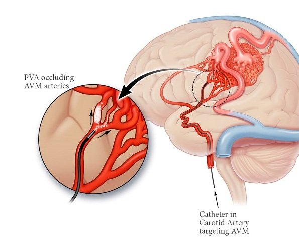
Treatment for brain AVMs can be symptomatic, and patients should be followed by a neurologist for any seizures, headaches, or focal neurologic deficits. AVM-specific treatment may also involve endovascular embolization, neurosurgery or radiosurgery.[2] Embolization, that is, cutting off the blood supply to the AVM with coils, particles, acrylates, or polymers introduced by a radiographically guided catheter, may be used in addition to neurosurgery or radiosurgery, but is rarely successful in isolation except in smaller AVMs.[15] Gamma knife may also be used.[16]
Treatment is offered is to try to prevent bleeding from the AVM. Bleeding may injure the surrounding brain resulting in a stroke , with possible permanent disability or even death. The risk of bleeding is 4% per year, which means that 4 out of every 100 people with an AVM will have a bleed (hemorrhage) during any one year. AVM’s may also produce headaches, seizures and progressive paralysis, and the treatment may alleviate these symptoms.
Your doctor will recommend the best treatment for you and this will be determined by the size of your AVM and also the location. It is not uncommon to recommend a combination of treatments.
Another option is to do nothing at all and just monitor the AVM. Your doctors may recommend observation if they feel that treatment can not be offered safely or when an AVM is discovered at a late age
Your treatment plan will depend on your age, condition, and physical health. The most important goal is to prevent internal bleeding, which can lead to stroke or death.
For more information visit us our website: https://www.healthinfi.com
0 200
No Comments