About Bilateral Temporal Lobe Hyperintensity (BTH) & Bilateral Temporal Lobe Disorder
Bilateral temporal lobe hyperintensity (BTH) is a commonly encountered MRI finding in a wide spectrum of clinical conditions and often poses a diagnostic challenge to the radiologist. The purpose of this paper is to elucidate several diseases that manifest as BTH on MRI, based on a retrospective review of cranial MRI of 65 cases seen in our institution between October 2007 and September 2010.
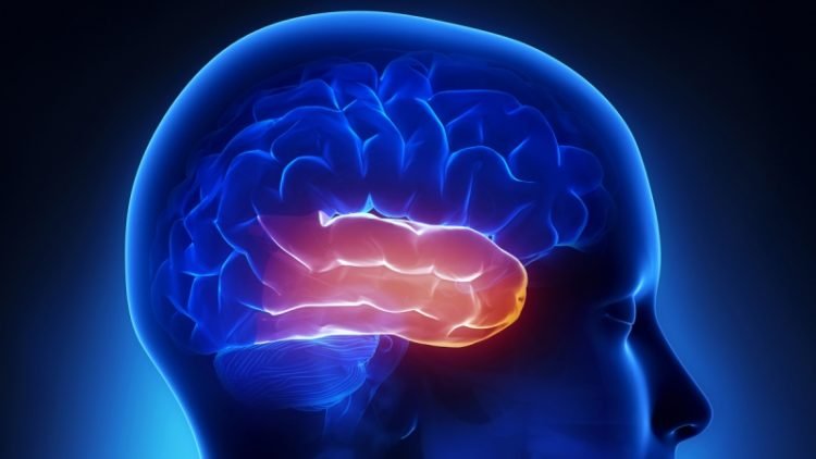
The temporal lobe is one of the four major lobes of the cerebral cortex in the brain of mammals. The temporal lobe is located beneath the lateral fissure on both cerebral hemispheres of the mammalian brain.
The temporal lobe is involved in processing sensory input into derived meanings for the appropriate retention of visual memory, language comprehension, and emotion association.
We found BTH in different clinical scenarios that included infective diseases (herpes simplex virus, congenital cytomegalovirus infection), epileptic syndrome (mesial temporal sclerosis), neurodegenerative disorders (Alzheimer’s disease, frontotemporal dementia, Type 1 myotonic dystrophy), neoplastic conditions (gliomatosis cerebri), metabolic disorders (mitochondrial encephalopathy, lactic acidosis and stroke-like episodes, Wilson’s disease, hyperammonemia), dysmyelinating disease (megalencephalic leukoencephalopathy with subcortical cysts), and vascular (cerebral autosomal dominant arteriopathy with subcortical infarcts and leukoencephalopathy) and paraneoplastic (limbic encephalitis) disorders.
")
Bilateral Temporal Lobe Hyperintensity (BTH)
The conventional MRI findings with advanced MRI such as diffusion-weighted imaging, susceptibility-weighted imaging and MR spectroscopy along with laboratory results are potentially helpful in distinguishing the different clinical conditions and thus affect the early diagnosis and clinical outcome.
The temporal lobes have unique architecture, and functionality that makes them vulnerable to certain disease processes. Patients presenting with bilateral temporal lobe disease are often confused and have altered consciousness, and are therefore unable to provide cogent histories. For these reasons, imaging plays an important role in their workup and management.
Disease entities causing bilateral temporal lobe involvement can be infectious, metabolic, neoplastic, and degenerative aetiologies, as well as trauma and cerebrovascular events. We will first describe the structural and functional anatomy of the temporal lobes and explain the mechanisms that underlie bilateral temporal lobe disease, and then show and discuss the different disease entities and differential diagnosis.
General Discussion
Klüver-Bucy syndrome is a very rare cerebral neurological disorder associated with damage to both temporal lobes resulting in abnormalities in memory, social and sexual functioning and idiosyncratic behaviors.
Temporal lobe anatomy
Functional centres of hearing, speech, memory, olfaction, sensation, emotion and behaviour are located in the temporal lobes, which lie in the middle cranial fossa, lateral to the midbrain and inferior to the basal ganglia. Each temporal lobe is separated from the frontal lobe and the anterior parietal lobe by the Sylvian fissure (Fig. 1). On axial images along the canthomeatal plane, the temporal lobes do not extend above the level of the lateral ventricles.
Signs & Symptoms
Major symptoms may include excessive oral tendencies with an urge to put all kinds of objects into the mouth, hypermetamorophosis (a need to explore everything), memory loss, emotional changes, extreme sexual behavior, indifference, placidity, visual distractibility and visual agnosia (difficulty identifying and processing visual information).
An almost uncontrollable appetite for food may also be noted. There may also be other symptoms associated with dementia (loss of reason) as well.
Aetiology
The most common cause of temporal lobe lesions is a CVE.
Space-occupying lesions may be primary brain tumours – benign (such as meningioma) or malignant. They may also be secondary tumours or metastatic carcinoma, most often from lung cancer or breast cancer.
Trauma from head injury may be involved or surgical damage when removing a tumour from that region. Head injury often includes extradural haematoma and contrecoup injuries (brain injury on the opposite side to the point of impact). Surgery for intractable temporal lobe epilepsy is well established and will cause disturbance of temporal lobe function.
Progressive deterioration of language can be part of a frontotemporal dementia. It presents earlier than Alzheimer’s disease and about 50% have a family history that suggests an autosomal dominant inheritance.
Presentation
A stroke tends to produce a rapid onset of symptoms whilst a space-occupying lesion will produce a more insidious onset. Whilst a hemiparesis is obvious to the patient and family (and will be recognised as such), the manifestations of temporal lobe lesions are more subtle and they may be interpreted as psychosis or dementia.
It is Important to unravel these strange presentations and to suspect the diagnosis. A careful, detailed history is required with examination. Often the patient will be oblivious to symptoms and will be uncomplaining. Some history from a third party can be useful.
There are eight principal symptoms of temporal lobe damage:
- Disturbance of auditory sensation and perception.
- Disturbance of selective attention of auditory and visual input.
- Disorders of visual perception.
- Impaired organisation and categorisation of verbal material.
- Disturbance of language comprehension.
- Impaired long-term memory.
- Altered personality and affective behaviour.
- Altered sexual behaviour.
- Manifestations of temporal lobe lesions
Causes
Klüver-Bucy syndrome is the result of damage to the temporal lobes of the brain. This may be the result of trauma to the brain itself, or the result of other degenerative brain diseases, tumors, or it can be caused by some brain infections, most commonly herpes simplex encephalitis (a viral brain infection).
Affected Populations
Klüver-Bucy syndrome is a very rare disease that affects males and females equally.
Related Disorders
Symptoms of the following disorders can be similar to those of Klüver-Bucy syndrome. Comparisons may be useful for a differential diagnosis:
Frontotemporal degeneration is a very rare progressive neurological disease initially predominately affecting the frontal and temporal lobes of the brain. It is characterized by progressive deterioration of intellect with changes in behavior and personality.
The memory is usually intact in the early stages of the disease and there is less disorientation than in Alzheimer’s disease. However, in later stages there is loss of motor control as well as confusion and severe dementia. (For more information on this disorder, choose “frontotemporal degeneration” as your search term in the Rare Disease Database.)
Alzheimer disease is a common progressive disorder of the brain affecting memory, thought and language. Groups of nerve endings in the cortex of the brains of people with Alzheimer’s degenerate and disrupt the passage of electrochemical signals between the cells.
Affected individuals become increasingly forgetful. As memory losses increase, personality, mood and behavior also tend to change. Judgment, concentration, speech and physical coordination may also be affected. (For more information on this disorder, choose “Alzheimer” as your search term in the Rare Disease Database.)
Korsakoff syndrome is a deficiency of vitamin B-1 that leads to cardiovascular, central and peripheral nervous system disturbances. Early symptoms of Korsakoff’s syndrome include fatigue, irritation, poor memory, difficulty sleeping, chest pain, abdominal discomfort, poor appetite and constipation.
Later symptoms are principally cardiovascular and neurological. (For more information on this disorder, choose “Korsakoff” as your search term in the Rare Disease Database.)
Visual memories
The temporal lobe communicates with the hippocampus and plays a key role in the formation of explicit long-term memory modulated by the amygdala.
Processing Sensory Input
Auditory
Adjacent areas in the superior, posterior, and lateral parts of the temporal lobes are involved in high-level auditory processing. The temporal lobe is involved in primary auditory perception, such as hearing, and holds the primary auditory cortex. The primary auditory cortex receives sensory information from the ears and secondary areas process the information into meaningful units such as speech and words.
The superior temporal gyrus includes an area (within the lateral fissure) where auditory signals from the cochlea first reach the cerebral cortex and are processed by the primary auditory cortex in the left temporal lobe.[citation needed]
Visual
The areas associated with vision in the temporal lobe interpret the meaning of visual stimuli and establish object recognition.[citation needed] The ventral part of the temporal cortices appear to be involved in high-level visual processing of complex stimuli such as faces (fusiform gyrus) and scenes (parahippocampal gyrus).[citation needed] Anterior parts of this ventral stream for visual processing are involved in object perception and recognition.
Language Recognition
The temporal lobe holds the primary auditory cortex, which is important for the processing of semantics in both speech and vision in humans. Wernicke’s area, which spans the region between temporal and parietal lobes, plays a key role (in tandem with Broca’s area in the frontal lobe) in speech comprehension. The functions of the left temporal lobe are not limited to low-level perception but extend to comprehension, naming, and verbal memory.[citation needed]
New memories
Emotion and memory
The medial temporal lobes (near the sagittal plane) are thought to be involved in encoding declarative long term memory.194–199 The medial temporal lobes include the hippocampi, which are essential for memory storage, therefore damage to this area can result in impairment in new memory formation leading to permanent or temporary anterograde amnesia.
Clinical significance
Unilateral temporal lesion
- Contralateral homonymous upper quadrantanopia (sector anopsia)
- Complex hallucinations (smell, sound, vision, memory)
Dominant hemisphere
- Receptive aphasia
- Wernicke’s aphasia
- Anomic aphasia
- Dyslexia
- Impaired verbal memory
- Word agnosia, word deafness
Non-dominant hemisphere
- Impaired non-verbal memory
- Impaired musical skills
Bitemporal lesions (additional features)
- Deafness
- Apathy (affective indifference)
- Impaired learning and memory
- Amnesia, Korsakoff syndrome,
Damage
Individuals who suffer from medial temporal lobe damage have a difficult time recalling visual stimuli. This neurotransmission deficit is due, not to lacking perception of visual stimuli but, to lacking perception of interpretation.
The most common symptom of inferior temporal lobe damage is visual agnosia, which involves impairment in the identification of familiar objects. Another less common type of inferior temporal lobe damage is prosopagnosia which is an impairment in the recognition of faces and distinction of unique individual facial features.
Damage specifically to the anterior portion of the left temporal lobe can cause savant syndrome.
Disorders
Pick’s disease, also known as frontotemporal amnesia, is caused by atrophy of the frontotemporal lobe.Emotional symptoms include mood changes, which the patient may be unaware of, including poor attention span and aggressive behavior towards themselves and/or others. Language symptoms include loss of speech, inability to read and/or write, loss of vocabulary and overall degeneration of motor ability.
Temporal lobe epilepsy is a chronic neurological condition characterized by recurrent seizures; symptoms include a variety of sensory (visual, auditory, olfactory, and gustation) hallucinations, as well as an inability to process semantic and episodic memories.
Schizophrenia is a severe psychotic disorder characterized by severe disorientation. Its most explicit symptom is the perception of external voices in the form of auditory hallucinations. The cause of such hallucinations has been attributed to deficits in the left temporal lobe, specifically within the primary auditory cortex.
Decreased gray matter, among other cellular deficits, contribute to spontaneous neural activity that affect the primary auditory cortex as if it were experiencing acoustic auditory input. The misrepresentation of speech in the auditory cortex results in the perception of external voices in the form of auditory hallucinations in schizophrenic patients. Structural and functional fMRI techniques have accounted for this neural activity by testing affected and non-affected individuals with external auditory stimuli.
Brain tissue processing
Paraformaldehyde (PFA)-fixed tissue
Rats were anaesthesized and euthanized by decapitation without tissue perfusion; brains were removed and kept for 10 days in 4% PFA at 4°C. Subsequently, brains were cryoprotected with 30% sucrose for 48 h, embedded in freezing medium, and snap-frozen in isopentane chilled with liquid nitrogen.
Other tissue processing
Brains from rats perfused with 4% PFA were removed and kept in 4% PFA for 1 h, and subsequently cryoprotected and embedded in freezing medium as above. Non-perfused rat brains were removed and directly embedded in freezing medium without fixative.
Immunoblot and immunohistochemistry
Sera (diluted 1 : 500) and CSF (1 : 10) were examined for antibodies using an immunoblot avidin–biotin peroxidase assay, as reported (Bataller et al., 2003). Immunoblots included protein extracts (100 µg/ml) from purified human cortical neurons, Purkinje cells and the recombinant proteins, HuD, Cdr2, Nova, Ma1, Ma2, CRMP5 and amphiphysin.
Immunohistochemistry was performed with cryostat-cut 7 µm thick sections mounted directly on slides. Non-pre-fixed tissue was incubated for 10 min with acetone or methanol–acetone at 4°C. Subsequently, all tissue sections were serially incubated with 0.25% H2O2 for 20 min, 10% goat serum for 30 min, the patient’s serum or CSF at the indicated dilutions in 10% goat serum overnight at 4°C, biotinylated goat anti-human IgG (1 : 2000) for 2 h and avidin–biotin peroxidase for 1 h, and the reactivity developed with diaminobenzidine.
Other primary antibodies used in consecutive tissue sections included: polyclonal rabbit antibodies to VGKCs Kv1.1, Kv1.2 and Kv1.6 (dilution 1 : 50; all from Sigma, St Louis, MO); a monoclonal antibody to Kv1.2 (1 : 50; Upstate Laboratories, Lake Placid, NY); a polyclonal antibody to synaptophysin (1 : 1000, Sigma); a monoclonal antibody to spinophilin (1 : 50; Upstate Laboratories); and human control serum with amphiphysin and Hu antibodies (1 : 500).
Intrathecal synthesis of antibodies was determined using serum and CSF samples normalized with the same concentration of IgG and serially diluted in parallel. Patients were considered to have intrathecal synthesis of antibodies if the end dilution point of CSF showed reactivity that was no longer present in the paired serum containing the same amount of IgG.
Immunocompetition assay
To determine whether patients’ antibodies targeted similar epitopes, immunocompetition assays were used, as reported (Dalmau et al., 1992). In brief, tissue sections pre-incubated with patient’s serum (or control normal serum) were subsequently incubated with IgG isolated from the serum of another patient and labelled with biotin. Abrogation of reactivity indicated that the patient’s serum contained antibodies similar to those present in the biotinylated IgG.
Double immunolabelling of hippocampal neurons
Hippocampal rat neuronal cell cultures were prepared as reported (Buchhalter and Dichter, 1991). Neurons were grown on coverslides, fixed with PFA, and serially incubated with serum from one patient (diluted 1 : 250) for 1 h, and goat anti-human IgG labelled with fluorescein for 30 min.
After washing, slides were incubated with biotinylated IgG from another patient with neuropil antibodies (or VGKC antibodies, or IgG from a normal individual), or the indicated monoclonal or polyclonal antibodies to VGKCs, spinophilin or synaptophysin for 1 h.
The reactivity of biotinylated human IgG was demonstrated with avidin–rhodamine for 30 min, and the reactivity of antibodies made in mouse or rabbit with the appropriate rhodamine-labelled secondary antibodies. Coverslides containing the neurons were mounted using aqueous medium.
0 200

No Comments