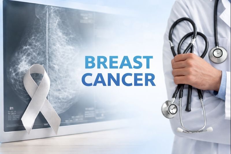
Breast cancer is a common cancer in women worldwide, but it can also affect men. Early detection plays a crucial role in improving survival rates, making awareness of breast cancer symptoms essential. Understanding the causes, risk factors, and available treatment options can help patients and caregivers make informed care decisions.
What Is Breast Cancer?
Cancer occurs when cells in the breast grow uncontrollably, forming a tumor. Tumors in the breast can be benign (non-cancerous) or malignant (cancerous). Malignant tumors can invade nearby tissues or spread to other areas of the body. Knowledge of cancer symptoms helps in early detection and timely medical intervention.
Breast Cancer Symptoms on Breast
Recognizing breast cancer symptoms is vital for early diagnosis. Common signs include:
-
A lump or thickening in the or underarm
-
Changes in breast size or shape
-
Skin dimpling or puckering
-
Nipple discharge, sometimes bloody
-
Redness, scaliness, or irritation of the skin
-
Pain or tenderness in the breast
Not all lumps are cancerous, but any unusual changes should be evaluated by a healthcare provider promptly.
Etiology for Breast Cancer
The etiology of cancer involves multiple factors, including genetic, hormonal imbalance, and environmental influences. Key elements include:
-
Genetic mutations: Mutations in BRCA1, BRCA2, or other genes increase risk.
-
Hormonal factors: Long-term exposure to estrogen and progesterone can influence cancer development.
-
Age and gender: Women over 50 are at higher risk, though younger females and males can also develop cancer.
-
Lifestyle factors: Obesity, alcohol consumption, smoking, and lack of physical activity may contribute.
Understanding the breast cancer reason behind each case often requires a combination of these factors.
Breast Cancer Risk Factors
Some individuals are at higher risk due to a combination of genetic, lifestyle, and reproductive factors. Common risk factors include:
-
Family history of cancer
-
Early menstruation or late menopause
-
Previous radiation exposure to the chest
-
Dense breast tissue
-
Hormone replacement therapy
Awareness of these factors can guide regular screenings and preventive strategies.
Breast Cancer Diagnosis
Early diagnosis of cancer significantly improves outcomes. Common diagnostic methods include:
-
Mammogram: X-ray imaging to detect early tumors
-
Ultrasound: Helps distinguish solid lumps from fluid-filled cysts
-
Biopsy: Removal of tissue for microscopic examination
-
MRI and genetic testing: Used in high-risk cases or complex diagnoses
Detecting breast cancer symptoms early is critical, as treatment is more effective in the initial stages.
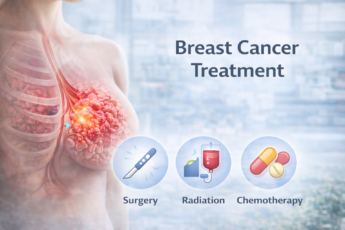
Breast Cancer Treatment
The cancer treatment depends on the stage, type, and the patient’s health. Common approaches include:
-
Surgery
-
Lumpectomy removes the tumor and some surrounding tissue.
-
Mastectomy involves removing one or both breasts.
-
-
Radiation Therapy
-
Used after surgery to destroy remaining cancer cells.
-
-
Chemotherapy
-
Uses medication to target and destroy rapidly dividing cells in the body.
-
-
Hormone Therapy
-
Blocks hormones like estrogen that fuel certain cancers.
-
-
Targeted Therapy
-
Focuses on specific molecules in cancer cells for precision treatment.
-
-
Immunotherapy
-
Strengthens the body’s immune system to fight cancer.
-
Treatment plans often combine multiple methods depending on individual needs, stage, and type of cancer, which is referred to as cancer therapy planning.
Lifestyle and Supportive Care
Alongside medical treatment, supportive care helps manage side effects and improve quality of life:
-
Healthy diet: A balanced diet supports recovery and overall health.
-
Physical activity: Gentle exercise reduces fatigue and improves mood.
-
Emotional support: Emotional support through counseling, support groups, and family can help manage stress.
-
Regular follow-up: Monitoring post-treatment is crucial for detecting recurrence early.
Patients should discuss all lifestyle interventions with their healthcare provider to ensure they complement the primary breast cancer treatment plan.
Preventive Measures
While not all cancer cases can be prevented, risk reduction strategies include:
-
Regular screenings such as mammograms
-
Maintaining a healthy weight
-
Limiting alcohol consumption
-
Avoiding tobacco use
-
Being aware of family history and considering genetic counseling
These measures help identify breast cancer symptoms early, which can significantly improve treatment outcomes.
Conclusion
Breast cancer is a complex disease with multiple causes and risk factors. Awareness of cancer symptoms and early detection are critical in improving survival rates. Understanding the etiology of cancer and the common cancer types helps individuals take proactive steps. Treatment options, including surgery, chemotherapy, radiation, hormone therapy, and newer targeted therapies, are tailored for each patient. Combining medical care with lifestyle changes and supportive therapy enhances overall well-being.
By recognizing early signs and consulting healthcare professionals promptly, patients can make informed decisions regarding cancer and therapy and access the best cancer treatment available.
FAQs
1. What are common cancer symptoms?
Lumps in the breast, changes in shape, nipple discharge, skin dimpling, or redness may indicate cancer.
2. What causes cancer?
The causes include genetic mutations, hormone imbalances, age, lifestyle factors, and family history—these make up the etiology for cancer.
3. How is cancer treated?
Treatment may include surgery, chemotherapy, radiation, hormone therapy, targeted therapy, or immunotherapy, depending on cancer type and stage.
4. Can lifestyle changes help with cancer recovery?
Yes, a healthy diet, exercise, stress management, and support systems complement medical treatment and improve overall well-being.
5. When is it necessary to see a doctor for changes in the breast?
Any unusual lump, persistent pain, nipple discharge, or skin changes in the breast should be evaluated immediately.
Reference
https://my.clevelandclinic.org/health/diseases/3986-breast-cancer</a>
https://www.cancer.gov/types/breast</a>
https://www.who.int/news-room/fact-sheets/detail/breast-cancer</a>
0 200
No Comments